When an electron beam drills holes in heated graphene, single-atom vacancies, shown in purple, diffuse until they join with other vacancies to form stationary structures and chains, shown in blue. Credit: Ondrej Dyck/ORNL, U.S. Dept. of Energy

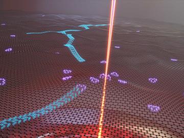
Oak Ridge National Laboratory researchers serendipitously discovered when they automated the beam of an electron microscope to precisely drill holes in the atomically thin lattice of graphene, the drilled holes closed up.

Bruce Warmack is using his physics and electrical engineering expertise to analyze advanced sensors for the power grid on a new testbed he developed at the Distributed Energy Communications and Controls Laboratory at ORNL. Credit: Carlos Jones/ORNL, U.S. Dept. of Energy

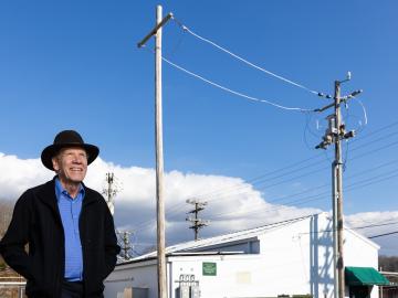
Bruce Warmack has been fascinated by science since his mother finally let him have a chemistry set at the age of nine. He’d been pestering her for one since he was six.

Colorized micrograph of lily pollen

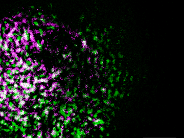
Oak Ridge National Laboratory researchers have built a novel microscope that provides a “chemical lens” for viewing biological systems including cell membranes and biofilms.

Materials — Molding molecular matter

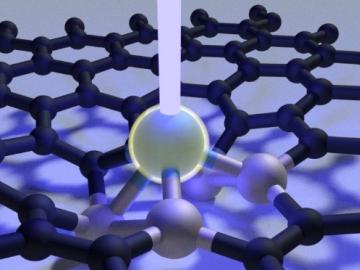
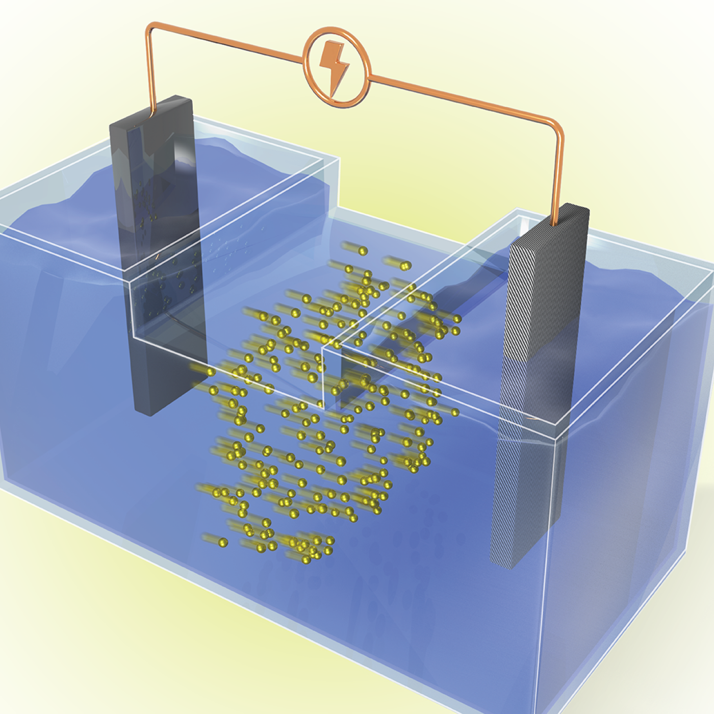
Scientists at Oak Ridge National Laboratory used a focused beam of electrons to stitch platinum-silicon molecules into graphene, marking the first deliberate insertion of artificial molecules into a graphene host matrix.

Scanning probe microscopes use an atom-sharp tip—only a few nanometers thick—to image materials on a nanometer length scale. The probe tip, invisible to the eye, is attached to a cantilever (pictured) that moves across material surfaces like the tone arm on a record player. Credit: Genevieve Martin/Oak Ridge National Laboratory; U.S. Dept. of Energy.

Liam Collins was drawn to study physics to understand “hidden things” and honed his expertise in microscopy so that he could bring them to light.