-
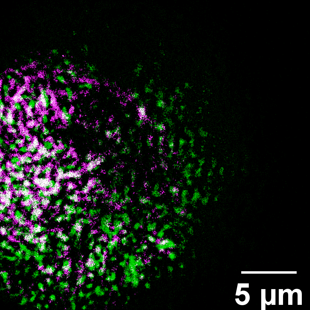
A novel ORNL microscope captured an image of lily pollen, which is colorized to show the distribution of two molecular groups. The instrument quickly shows chemical details. Credit: Uvinduni Premadasa/ORNL, U.S. Dept. of Energy
-
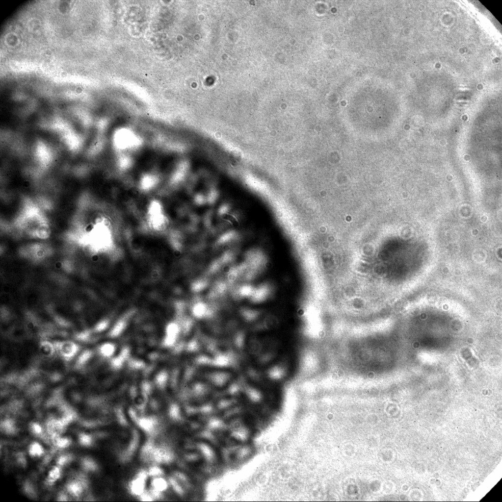
An image of lily pollen, captured using bright-field microscopy developed by ORNL, reveals only the presence of material without information about its composition. Credit: Uvinduni Premadasa/ORNL, U.S. Dept. of Energy
-
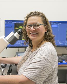
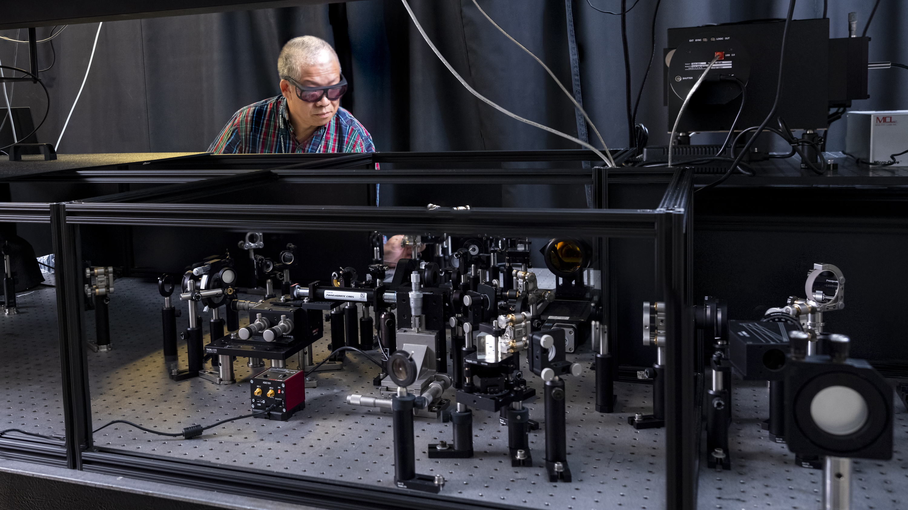
ORNL’s Yingzhong Ma examines a novel microscope that he built with collaborators. The microscope captures images using intense, ultrashort laser pulses. Credit: Carlos Jones/ORNL, U.S. Dept. of Energy
-
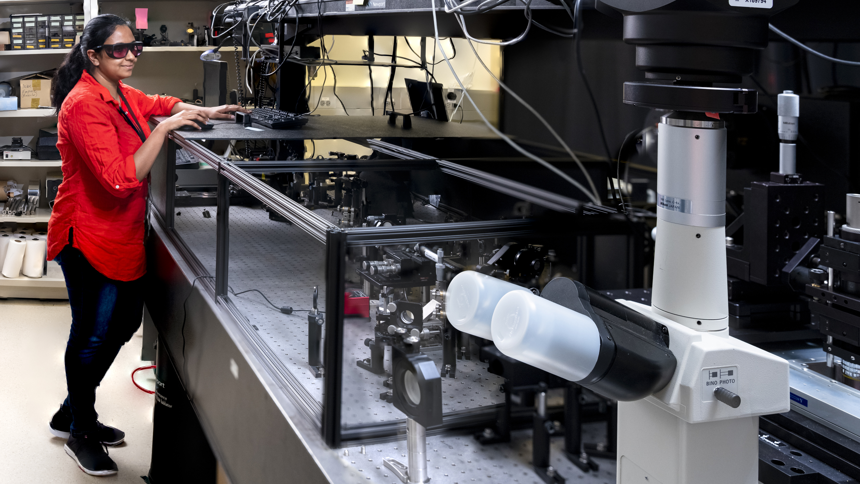
Postdoctoral research associate Uvinduni Premadasa stands beside the microscopy tool, which captures wide-field images of biological samples with chemical detail. Credit: Carlos Jones/ORNL, U.S. Dept. of Energy
-
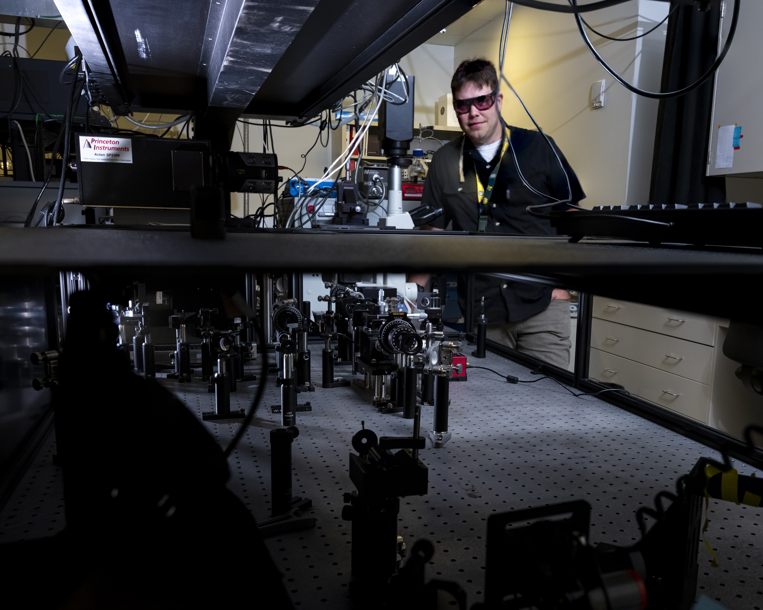
ORNL’s Benjamin Doughty stands behind the laser-enabled microscope. Credit: Carlos Jones/ORNL, U.S. Dept. of Energy
-
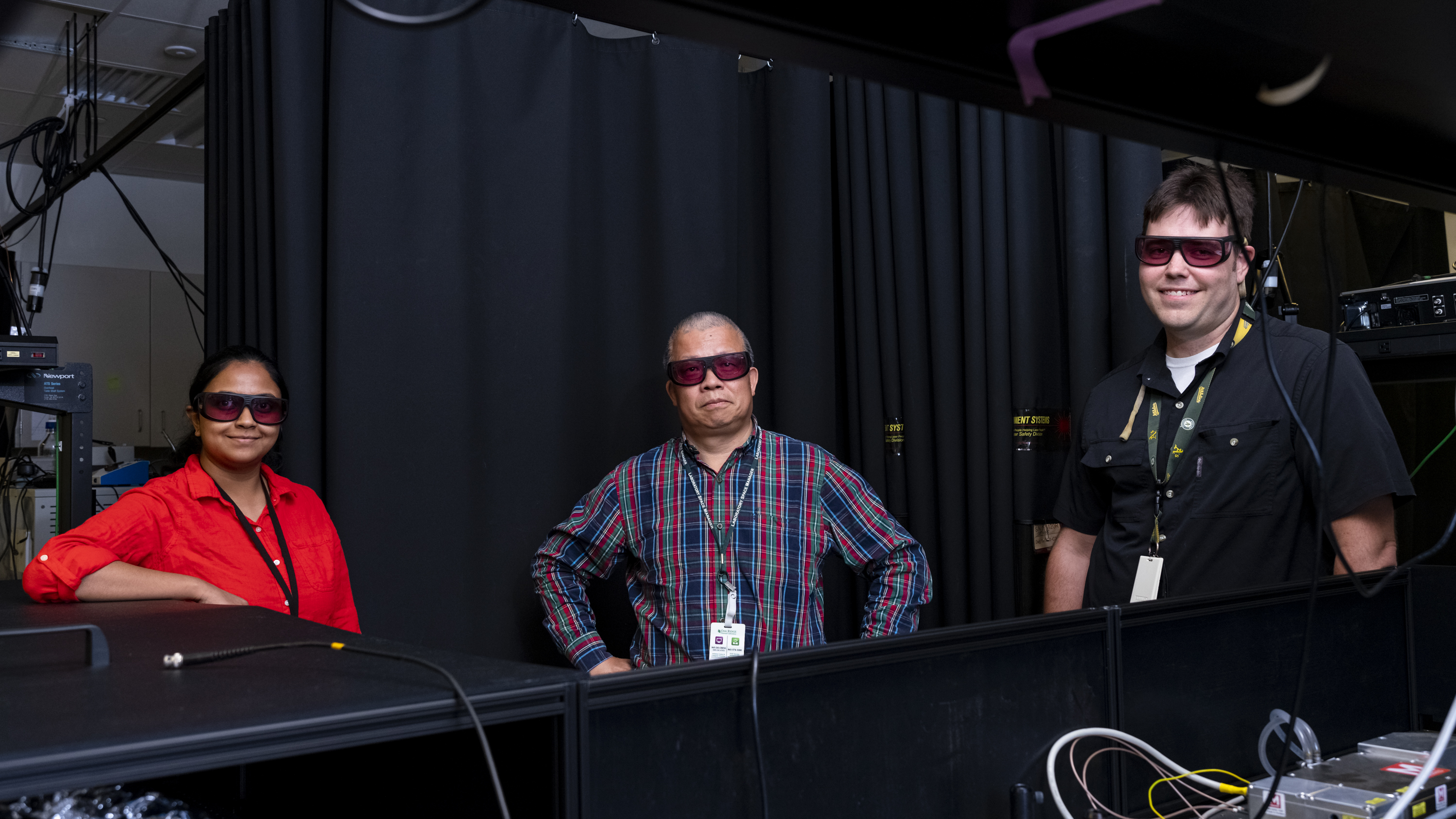
Uvinduni Premadasa, Yingzhong Ma and Benjamin Doughty developed a novel microscope for bioimaging. The tool was reported in the journal Optics Letters. Credit: Carlos Jones/ORNL, U.S. Dept. of Energy


-
A novel ORNL microscope captured an image of lily pollen, which is colorized to show the distribution of two molecular groups. The instrument quickly shows chemical details. Credit: Uvinduni Premadasa/ORNL, U.S. Dept. of Energy
-
An image of lily pollen, captured using bright-field microscopy developed by ORNL, reveals only the presence of material without information about its composition. Credit: Uvinduni Premadasa/ORNL, U.S. Dept. of Energy
-
ORNL’s Yingzhong Ma examines a novel microscope that he built with collaborators. The microscope captures images using intense, ultrashort laser pulses. Credit: Carlos Jones/ORNL, U.S. Dept. of Energy
-
Postdoctoral research associate Uvinduni Premadasa stands beside the microscopy tool, which captures wide-field images of biological samples with chemical detail. Credit: Carlos Jones/ORNL, U.S. Dept. of Energy
-
ORNL’s Benjamin Doughty stands behind the laser-enabled microscope. Credit: Carlos Jones/ORNL, U.S. Dept. of Energy
-
Uvinduni Premadasa, Yingzhong Ma and Benjamin Doughty developed a novel microscope for bioimaging. The tool was reported in the journal Optics Letters. Credit: Carlos Jones/ORNL, U.S. Dept. of Energy
Oak Ridge National Laboratory researchers have built a novel microscope that provides a “chemical lens” for viewing biological systems including cell membranes and biofilms. The tool could advance the understanding of complex biological interactions, such as those between microbes and plants.
The noninvasive instrument, detailed in Optics Letters, allows researchers to capture images using ultrashort laser pulses. These intense pulses illuminate large areas of a sample, generating colors of light that allow detection of different chemical species. The approach quickly produces images over a wide field of view with chemical details.
“Because you’re getting the whole image all in the same shot, you’re able to study changes in space and in time,” ORNL’s Benjamin Doughty said.
Unlike common bioimaging techniques that can destroy or disturb samples, this label-free tool can be used on unaltered, living cells. The microscope is made with commonly available components, which may accelerate its implementation.


