ORNL's Communications team works with news media seeking information about the laboratory. Media may use the resources listed below or send questions to news@ornl.gov.
1 - 9 of 9 Results

Scientists at ORNL and the University of Cincinnati achieved a breakthrough in understanding the vulnerability of microbes to the butanol they produce during fermentation of plant biomass. The discovery could pave the way for more efficient production of domestic fuels, chemicals and materials.

To help reduce the likelihood of losing future cultivated crops to drought and other seasonal hardships, researchers from ORNL, Budapest and Hungary are using neutrons, light microscopy and transmission electron microscopy to study the 'Never Never' plant, known for its ability to endure periods of little to no rain.

During his first visit to Oak Ridge National Laboratory, Energy Secretary Chris Wright compared the urgency of the Lab’s World War II beginnings to today’s global race to lead in artificial intelligence, calling for a “Manhattan Project 2.”
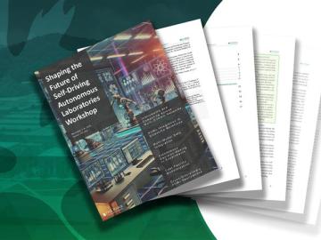
A workshop led by scientists at ORNL sketched a road map toward a longtime goal: development of autonomous, or self-driving, next-generation research laboratories.
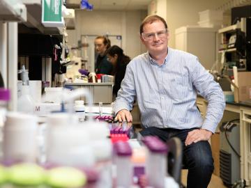
Hugh O’Neill’s lifelong fascination with the complexities of the natural world drives his research at ORNL, where he’s using powerful neutron beams to dive deep into the microscopic realm of biological materials and unlock secrets for better production of domestic biofuels and bioproducts.

A team of scientists led by a professor from Duke University discovered a way to help make batteries safer, charge faster and last longer. They relied on neutrons at ORNL to understand at the atomic scale how lithium moves in lithium phosphorus sulfur chloride, a promising new type of solid-state battery material known as a superionic compound.

Bio-SANS, the Biological Small-Angle Neutron Scattering Instrument at HFIR recently had a detector upgrade that will provide significantly improved performance that is more in line with the instrument’s capability.
We now know that many serious diseases have genetic links that a geneticist can find by reading an individual’s genome─the DNA double helix where our organism’s hereditary information is encoded. Researchers know too that a particular protein protects our DNA, which is vulnerable to entanglement when its information is read and to attack from enzymes that damage the strands, making the code indecipherable.
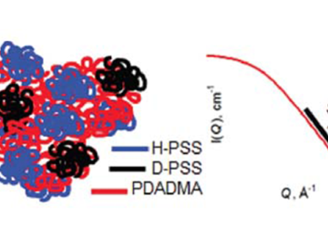
Researchers at the Bio-SANS instrument at the High Flux Isotope Reactor (HFIR) used small-angle neutron scattering (SANS) to get a first insight into the conformation of single polyelectrolyte chains in large pieces of the synthetic complex. The research pursues applications for replacement of intervertebral discs in the spine and of knee cartilage.