The first neutron structure of the SARS-CoV-2 main protease enzyme revealed unexpected electrical charges in the amino acids cysteine (negative) and histidine (positive), providing key data about the virus’s replication. Credit: Jill Hemman/ORNL, U.S. Dept. of Energy

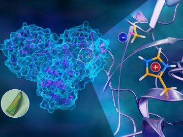
To better understand how the novel coronavirus behaves and how it can be stopped, scientists have completed a three-dimensional map that reveals the location of every atom in an enzyme molecule critical to SARS-CoV-2 reproduction.

ORNL researchers and energy storage startup Sparkz have developed a cobalt-free cathode material for use in lithium-ion batteries Credit: Ilias Belharouak/Oak Ridge National Laboratory, U.S. Dept. of Energy

Four research teams from the Department of Energy’s Oak Ridge National Laboratory and their technologies have received 2020 R&D 100 Awards.

Sergei Kalinin

Five researchers at the Department of Energy’s Oak Ridge National Laboratory have been named ORNL Corporate Fellows in recognition of significant career accomplishments and continued leadership in their scientific fields.

The protease protein is both shaped like a heart and functions as one, allowing the virus replicate and spread. Inhibiting the protease would block virus reproduction. Credit: Andrey Kovalevsky/ORNL, U.S. Dept. of Energy

A team of researchers has performed the first room-temperature X-ray measurements on the SARS-CoV-2 main protease — the enzyme that enables the virus to reproduce.

A nanobrush made by pulsed laser deposition of CeO2 and Y2O3 with dim and bright bands, respectively, is seen in cross-section with scanning transmission electron microscopy. Credit: Oak Ridge National Laboratory, U.S. Dept. of Energy

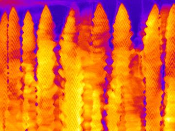
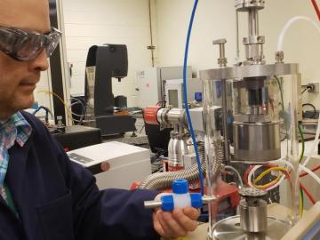
A team led by the Department of Energy’s Oak Ridge National Laboratory synthesized a tiny structure with high surface area and discovered how its unique architecture drives ions across interfaces to transport energy or information.

Illustration of the intricate organization of the PKA structure, wherein different parts of the protein are connected through elaborate hydrogen bonding networks (dashed yellow lines), glued together by the hydrophobic assemblies (light blue and orange volumes)—all working together to build the functional active site. Insert shows protonation of the transferred phosphoryl group (cyan mesh) and its many interactions with water and the active site amino acid residues. Credit: Jill Hemman/ORNL

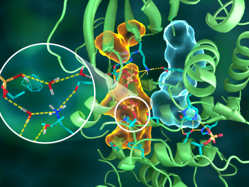
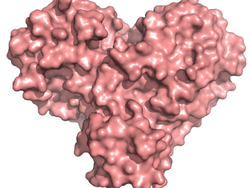
OAK RIDGE, Tenn., March 20, 2019—Direct observations of the structure and catalytic mechanism of a prototypical kinase enzyme—protein kinase A or PKA—will provide researchers and drug developers with significantly enhanced abilities to understand and treat fatal diseases and neurological disorders such as cancer, diabetes, and cystic fibrosis.